


Ellipse: Do you know the orbit of planets, moon, comets, and other heavenly bodies are elliptical? Mathematics defines an ellipse as a plane curve surrounding...
Last Modified 14-04-2025

Harvest Smarter Results!
Celebrate Baisakhi with smarter learning and steady progress.
Unlock discounts on all plans and grow your way to success!

Ellipse: Definition, Properties, Applications, Equation, Formulas
April 14, 2025
Altitude of a Triangle: Definition & Applications
April 14, 2025
Manufacturing of Sulphuric Acid by Contact Process
April 13, 2025
Refining or Purification of Impure Metals
April 13, 2025
Pollination and Outbreeding Devices: Definition, Types, Pollen Pistil Interaction
April 13, 2025
Electrochemical Principles of Metallurgy: Processes, Types & Examples
April 13, 2025
Acid Rain: Causes, Effects
April 10, 2025
Congruence of Triangles: Definition, Properties, Rules for Congruence
April 8, 2025
Complementary and Supplementary Angles: Definition, Examples
April 8, 2025
Nitro Compounds: Types, Synthesis, Properties and Uses
April 8, 2025
Homeostasis and Micturition: Homeostasis (“steady-state”) is a process by which animal organs and organ systems constantly respond to internal and external changes. Alterations in blood glucose or calcium levels, as well as external temperatures, could cause these changes. The term “homeostasis” refers to the body’s dynamic equilibrium.
It’s dynamic because it’s constantly adapting to the changes that the body’s systems go through. Because physiological functions are controlled within precise parameters, it is balanced. Micturition, also called urination, is the process of releasing urine from the bladder. Urination is used to remove metabolic products and harmful wastes from the body that the kidneys have filtered from the blood. Let’s take a deeper look at Homeostasis and Micturition, definition, and mechanism.
Homeostasis is critical for an organism’s survival. It is a self-regulatory process that maintains life by controlling internal factors. Homeostasis is a system that keeps the internal environment consistent despite changes in the external environment. Body temperature, blood pH, blood glucose levels, fluid balance, sodium, potassium, and calcium ion concentrations are all controlled by the body to maintain Homeostasis.

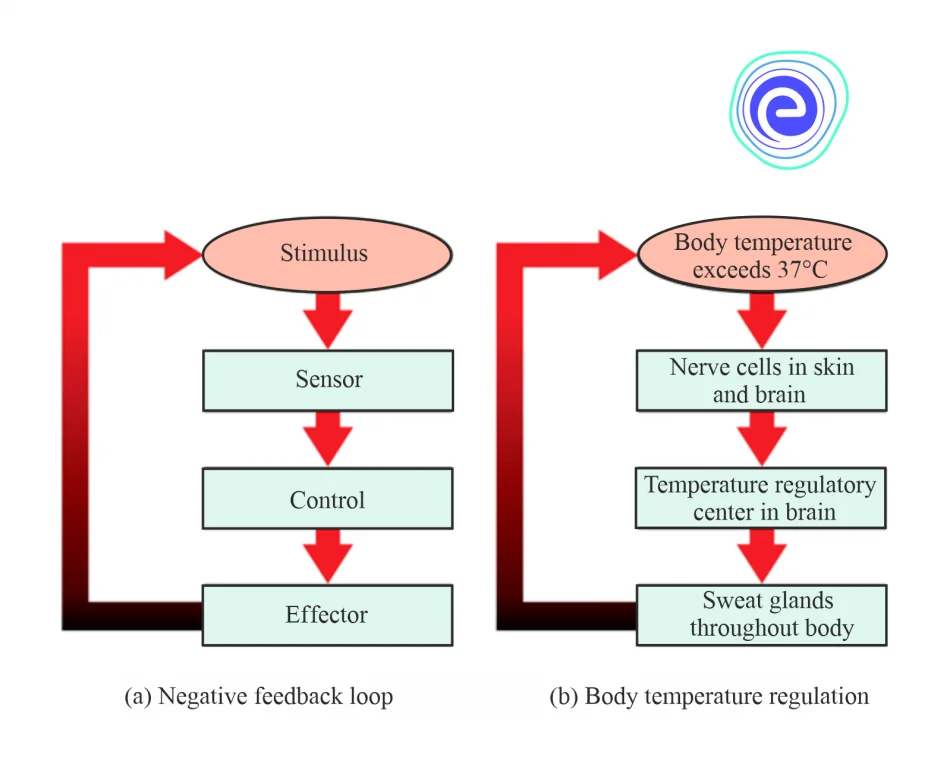
Fig: Regulation of Homeostasis
Three mechanisms are involved in maintaining homeostasis:
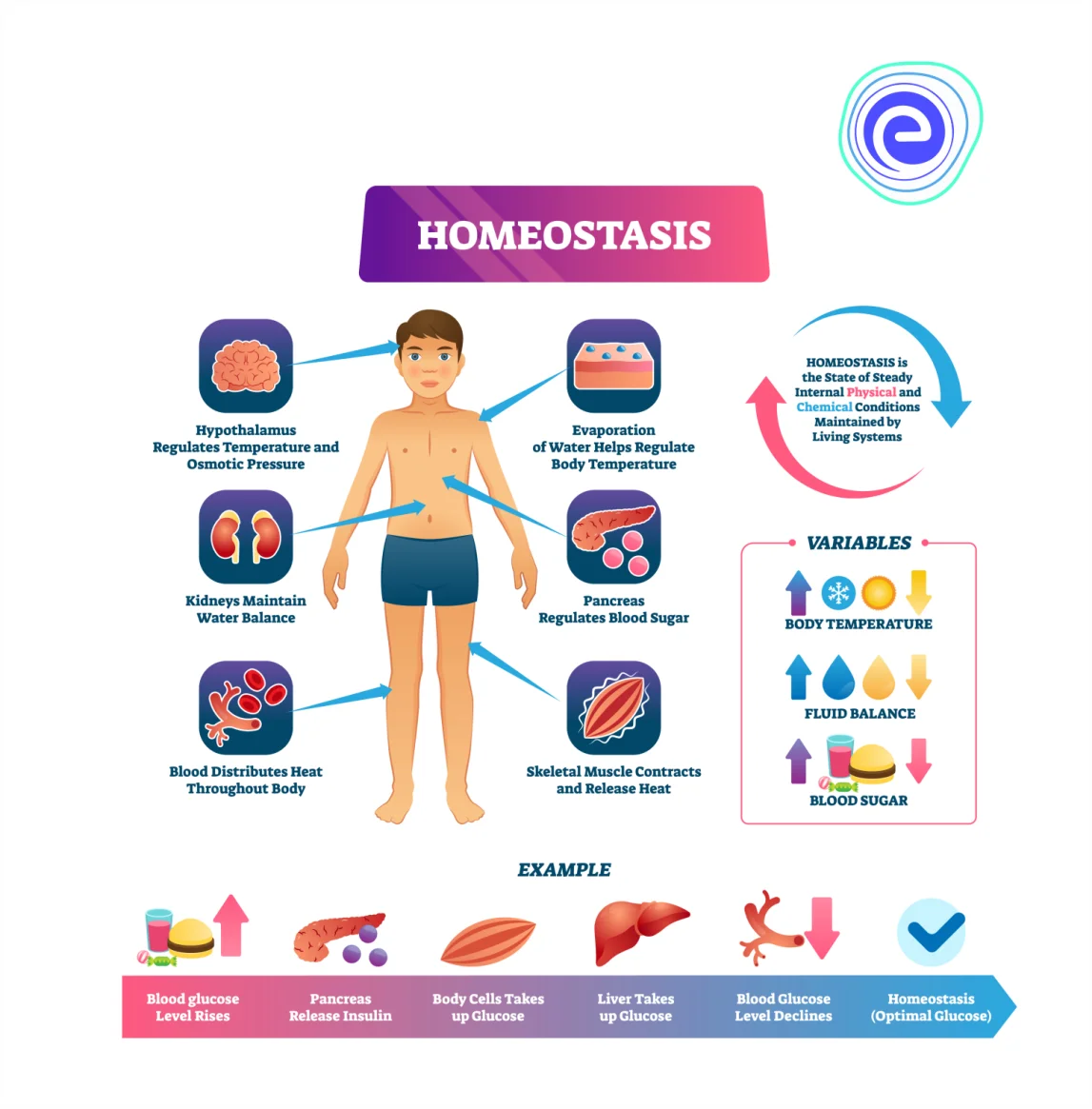
Fig: Homeostasis of Human Body
The purpose of Homeostasis is to keep things in balance around a specific point or value called a set point. While there will be typical variations from the setpoint, the body’s systems will normally try to return to it. A stimulus is a change in the receptor’s internal or external surroundings. The system adjusts the deviation parameter to bring it closer to the specified point. If the animal’s body becomes too hot, changes are made to cool it down. If blood glucose levels rise after a meal, changes are made to lower blood glucose levels by delivering nutrients to tissues that require them or storing them for later use.
A change in an animal’s environment necessitates a change in behaviour. The receptor detects a change in the environment and sends a signal to the control centre (usually the brain), which develops a response and signals it to an effector. A muscle (that contracts or relaxes) or a gland that secretes is the effector. Negative feedback loops keep homeostasis in action. Positive feedback loops push the organism further away from homeostasis, although they may be required for life to exist. The nervous and endocrine systems of mammals regulate homeostasis.
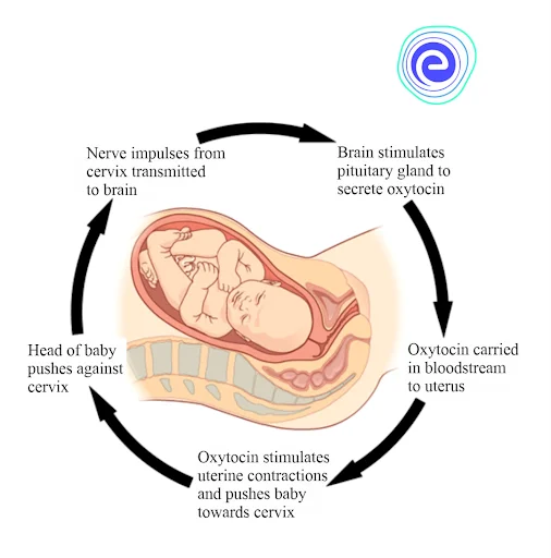
Fig: Positive Feedback Loops
The body’s activities are influenced by its temperature. In general, as body temperature rises, so does enzyme activity. Enzyme activity doubles for every \(10^\circ \) Celsius increase in temperature, up to a point. High heat causes body proteins, including enzymes, to denature and lose their activity (around \(50^\circ {\text{C}}\) for mammals). With a few exceptions, enzyme activity decreases by \(50\% \) for every ten degrees Celsius drop in temperature until the point ofzing. Some fish can tolerate a completeze and then thaw back to normal.
Animals can be divided into two groups:
The process of expelling urine from the bladder is known as micturition. Urination is another name for it. Urination is the process of removing metabolic products and toxic wastes from the body that have been filtered from the blood by the kidneys. It’s a sophisticated process combining the sympathetic, parasympathetic, and somatic nervous systems, and the brain’s higher centres allow urinating at the right time. Other crucial aspects in this process include normal muscular tone, the lack of physical blockages, and psychological inhibition.
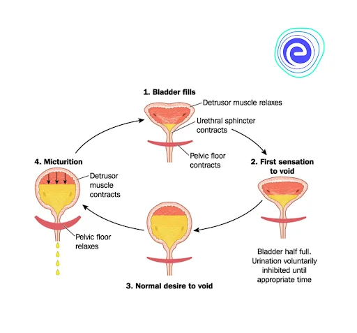
Fig: Micturition

There are two main stages or phases in the urinary bladder:
1. Resting or Filling Stage
a. Urine is carried from the kidneys to the bladder via the ureters during this phase of the bladder. The ureters are narrow muscular tubes that emerge from each kidney and extend downward, where they obliquely enter the bladder.
b. The ureters’ oblique positioning in the bladder wall provides a critical purpose. There are no sphincters or muscles to protect the entry of the ureter into the urine bladder. As a result of the oblique orientation of the incision, urine cannot re-enter the ureters. At the same time, the detrusor muscle, the main muscle of the urinary bladder, relaxes, allowing the bladder to expand and accommodate more urine.
2. Voiding Stage
a. Both the urine bladder and the urethra are involved during this stage. When the bladder’s storage capacity is achieved, the detrusor muscle of the urine bladder, which had been relaxing, begins to contract.
b. The internal and external urethral sphincters are two muscles that govern the urethra. The internal sphincter is made up of smooth muscles, whereas the external sphincter is made up of skeletal muscles. During the filling stage, both of these sphincters are contracted.
c. Urine passing is controlled by the parasympathetic nervous system. Bladder afferents send signals to the pontine micturition centre and the cerebrum after ascending through the spinal cord. The sacral preganglionic neurons are excited when neurons in the pontine micturition centre fire in response to a voluntary decision to urinate.
d. Following parasympathetic stimulation of the pelvic nerve (nerve roots S2-4), acetylcholine (ACh) is released, which acts on muscarinic ACh receptors (M3 receptors) on the detrusor muscle, causing it to contract and raise intravesicular pressure. The pontine micturition centre also inhibits Onuf’s nucleus, causing relaxation by reducing sympathetic input to the internal urethral sphincter.
e. Finally, a conscious reduction in the external urethral sphincter’s voluntary contraction from the cerebral cortex allows for urethral distention and urine passage.
f. The female’s urination is facilitated by gravity, but the male’s urination is facilitated by bulbospongiosus muscular contractions down the length of the penis.
The process by which animal organs and organ systems constantly adjust to internal and external changes is known as Homeostasis (“steady state”). These fluctuations could be caused by changes in blood glucose or calcium levels, as well as external temperatures. The dynamic equilibrium of the body is referred to as “Homeostasis.” It’s dynamic since it’s always responding to the changes that occur in the body’s systems. It is balanced because physiological functions are controlled within exact parameters.
Homeostasis involves the receptor, the control centre, and the effector. The information received by the receptor, which includes information about the changing environment, is processed by the control centre. The effector enhances or opposes the stimulus in response to the control centre’s directives. The process of discharging urine from the bladder is known as micturition or urination. Urination is the process of removing metabolic products and toxic wastes from the body that have been filtered from the blood by the kidneys.

Q.1. State homeostasis definition.
Ans: Homeostasis (“steady-state”) is a process by which animal organs and organ systems constantly respond to internal and external changes.
Q.2. Which body systems help to maintain homeostasis?
Ans: The neurological system and the endocrine system are both important in maintaining body homeostasis. Other organs, on the other hand, play a part in maintaining homeostasis.
Q.3. What is micturition?
Ans: The process of urine excretion from the urinary bladder is known as micturition.
Q.4. What are the main components of homeostasis?
Ans: Homeostasis involves three components- the receptor, the control centre, and the effector. The receptor receives information on the changing environment, and the control centre processes the information received by the receptor. The effector responds to the commands of the control centre by enhancing or opposing the stimulus.
Q.5. What is the storage phase and voiding phase?
Ans: The storage phase is characterised by the storage of urine by the urinary bladder. The movement is controlled by the circular sphincter muscles. The voiding phase is said to occur when the brain sends signals to begin urinating until the bladder becomes empty.
Learn About Kingdom Animalia Here
We hope this detailed article on Homeostasis and Micturition is helpful to you. If you have any queries, drop a comment below, and we will get back to you.


