


Ellipse: Do you know the orbit of planets, moon, comets, and other heavenly bodies are elliptical? Mathematics defines an ellipse as a plane curve surrounding...
Last Modified 14-04-2025

Harvest Smarter Results!
Celebrate Baisakhi with smarter learning and steady progress.
Unlock discounts on all plans and grow your way to success!

Ellipse: Definition, Properties, Applications, Equation, Formulas
April 14, 2025
Altitude of a Triangle: Definition & Applications
April 14, 2025
Manufacturing of Sulphuric Acid by Contact Process
April 13, 2025
Refining or Purification of Impure Metals
April 13, 2025
Pollination and Outbreeding Devices: Definition, Types, Pollen Pistil Interaction
April 13, 2025
Acid Rain: Causes, Effects
April 10, 2025
Congruence of Triangles: Definition, Properties, Rules for Congruence
April 8, 2025
Complementary and Supplementary Angles: Definition, Examples
April 8, 2025
Nitro Compounds: Types, Synthesis, Properties and Uses
April 8, 2025
Bond Linking Monomers in Polymers: Biomolecules, Diagrams
April 8, 2025
The Brain: Have you ever wondered how we think and react to the postures and gestures of the opponent? Which organ is responsible for thinking, responding, imagining things, etc. Yes, the brain is the important organ that has the ability to respond to stimuli. The brain is the organ that controls and coordinates the different systems of the body and weighs about 3 pounds in the average adult. All living organisms have a basic need to control their systems.
The brain is one of the largest and most complex organs in the human body and is made up of billions of neurons (or nerve cells) that communicate in trillions of connections called synapses. An electroencephalogram (EEG) records the waves of the brain. It is a medical test used to measure the electrical activity of the brain, and it is also used to detect abnormalities in brain waves. Read on more to know about the brain, parts of the brain, and function of the brain in the following article.

The brain is the most complex and important organ in the human body. It is a part of the nervous system that is responsible for many sites like thinking, imagination, intelligence, body movement, senses, behaviour control, etc. The nervous system in humans is divided into the central nervous system and peripheral nervous system, and the brain, along with the spinal cord, constitutes the central nervous system.
Fig: The Brain
Study About Human Body in Detail
The brain looks crescent-shaped inside the skull. The normal adult human brain weighs from 1300 to 1400 grams or around 3 pounds. In terms of measuring length, the brain is about 15 cm long. In general, the man’s brain weighs about 100 grams larger than the woman’s.
The brain weighs approximately 1400 g that is present inside the skull. The skull consists of 22 bones, of which 14 are facial bones, and the remaining 8 are cranial bones. The skull is also responsible for providing dorsal, frontal, and lateral protection to the brain. The brain is protected by the cranial bones and the meninges.
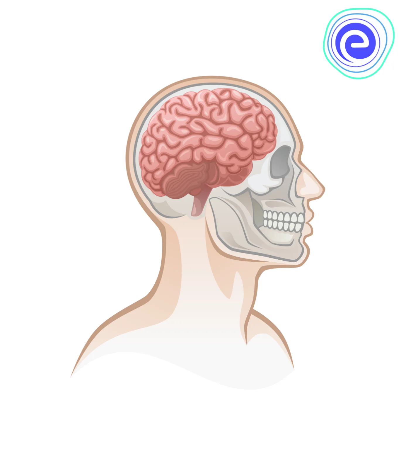
Fig: Location of the Brain
The whole central nervous system is further protected by the meninges, which are continuous with the spinal cord, consisting of three layers: the outermost dura mater, middle arachnoid, and the innermost pia mater.
The brain has the following divisions:
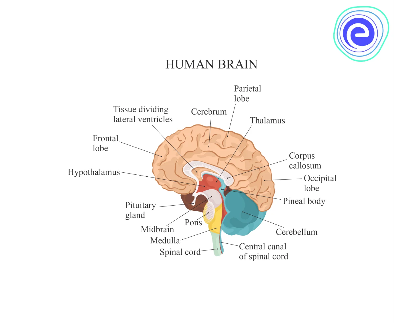
Fig: Parts of the Brain
It consists of the cerebrum and diencephalon.
1. Cerebrum: It is the largest part of the brain and is divided into right and left halves called cerebral hemispheres. These are connected together by a sheet of nerve fibres called the corpus callosum. The cerebrum is further subdivided into four sections or parts, and they are:

Functions of Olfactory Lobes: These are a pair of small club-shaped solid structures lying on the lower surface of cerebral hemispheres. Olfactory lobes are connected with the relay of sense of smell.
The wall of the cerebrum has two regions. The outer region is called the cortex and contains cell bodies of the neurons and is thus greyish in colour. So it is also called the grey matter. The grey matter is highly convoluted with ridges and grooves. These convolutions increase the surface area and total grey matter. The number of convolutions is believed to be associated with the degree of intelligence. The inner region of the cerebrum has white matter, which is composed of axons of the neurons.
Functions of Cerebrum: Seat of mental abilities, controls thinking, memory, reasoning, perception, emotions, and speech. Interprets sensations and responds to pain, cold, heat, and pressure.
2. Diencephalon: It lies concealed by the cerebrum. The diencephalon is distinguishable in two parts, the thalamus, and the hypothalamus.
(i) Thalamus is concerned with relaying sensory impulses except those of smell and regulation of smooth muscle activity.
(ii) Hypothalamus coordinates various autonomic activities of the body, including water balance, control of pituitary gland, body temperature, blood pressure, etc. Diencephalon has a cavity called the third ventricle. The roof of the diencephalon possesses an anterior choroid plexus for the formation of cerebrospinal fluid. The inferior surface of the diencephalon bears optic chiasma and the pituitary gland.
Functions of Diencephalon: Relay centre for sensory impulses, such as pain, temperature, and light.
1. Reflex centre for muscular activities.
2. Centre for certain emotions such as anger.
3. Centre for water balance, blood pressure, body temperature, sleep, and hunger.
4. It controls the pituitary gland, which functions as the master gland, body temperature, blood pressure, etc. Hypothalamus strongly influences water preservation and parturition.
5. The fibres from the hypothalamus also control the brainstem autonomic centres, particularly the vasomotor centre (VMC).
The midbrain is significantly small and consists of two heavy fibre tracts, known as cerebral peduncles (crura cerebri), on the ventral side. There are two swellings, superior and inferior colliculi, on each side of the dorsal surface. The midbrain connects the forebrain to the hindbrain and controls muscle toning, modifies some motor activities, and also has reflexes for sight and hearing.
Functions of the Midbrain: It relays motor impulses from the cerebral cortex to the pons and spinal cord and relays sensory impulses from the spinal cord to the thalamus.
The hindbrain consists of the cerebellum, pons, and medulla oblongata.
1. Cerebellum: It is very well-developed and lies below the cerebrum and above the medulla. It is known as a small brain. It consists of three parts – A middle vermis and two lateral hemispheres. The two hemispheres possess numerous furrows but no convolutions. Pons varoli is a bridge of nerve fibres connecting the two lobes of the cerebellum.
(i) Transmission of impulses across the bridge ensures coordination of muscular movements on both sides of the body. It has an outer cortex made of grey matter, while white matter is located centrally. The latter appears like a branching tree. Its fibre tracts are connected with the cerebrum and medulla oblongata.
(ii) This region is a motor area of the brain concerned with coordinating movements of skeletal muscles. It maintains posture, equilibrium, and muscular toning. Alcohol affects the coordination of the head, eyes, and limbs. A drunken driver does not have complete control over the vehicle because, under the influence of alcohol, the cerebellum cannot coordinate muscular movements.
Functions of the Cerebellum: It maintains posture, equilibrium, and muscle tone. Coordinates voluntary movements are initiated by the cerebrum. It mainly controls the balance and posture of the body. Its control is ipsilateral over half of the body on the same side.
2. Pons: On the ventral side of the brain, below the cerebellum, a thick bundle of fibres is present that is known as pons varoli. It transmits impulses from one side of the cerebellum to the other, and together with the medulla, it controls breathing.
Functions of Pons: Transmits impulses from one side of the cerebellum to the other, and together with the medulla, it controls breathing. Pons control the sleep cycles.
3. Medulla Oblongata: It is the lowest part of the brain located at the base of the skull. It is conical in outline and continues below with the spinal cord. It has white matter on the outer side and grey matter on the inner side. Medulla oblongata, pons varoli, midbrain, and hypothalamus are collectively known as the brainstem. They are hidden by the cerebrum and cerebellum.
(i) The roof of the medulla oblongata contains a choroid plexus where cerebrospinal fluid is formed. The posterior medulla controls the activities of the internal organs. It has reflex centres such as cardiac centres, respiratory centres and vasomotor centres.
Functions of the Medulla Oblongata: Contains centre for cardiac, respiratory, and vasomotor activities. Coordinates reflexes like swallowing, coughing, sneezing, and vomiting.
Study Human Body and Movements
There are mainly four ventricles of the brain. Lateral ventricles are the first two ventricles that are connected to the third ventricle through a prominent opening called the Foramen of Munro. The communication between the third and the fourth ventricle takes place via the Aqueduct of Silvius that is a long tube.
1. Lateral Ventricle: The first and second combined together is called the lateral ventricle; they are enclosed in the cerebral hemispheres.
2. Third Ventricle: It is present in the centre of the brain, and its walls are made up of the hypothalamus and thalamus. The pineal gland is situated behind the third ventricle. It helps to control the inner clock and circadian rhythms of the body through the secretion of melatonin.
3. Fourth Ventricle: The fourth ventricle is located in the posterior or dorsal to the pons and medulla oblongata and is anterior to the cerebellum.
The nervous system is a very important fortune to the body because of the vital role it plays in normal functioning. The brain is the control tower of the human body. It helps in controlling and coordinating the actions and reactions that are received from the surroundings. The brain is divided into three main parts- forebrain, midbrain, and hindbrain.
The forebrain is the largest part of the brain that contains major parts called the cerebrum, hypothalamus, thalamus. The midbrain is the smallest and central part of the brain that consists of two divisions called tectum and tegmentum. The last part of the brain is the hindbrain. It is the lowest part of the brain that is composed of the cerebellum, pons, and medulla. The brain has the capacity to control all the actions like walking, sleeping, breathing, body temperature, emotions, etc.

Ans: The brain is the main and important organ of the human body because it controls and coordinates all the actions and reactions of the body. It is the organ responsible for thinking, feeling, imagining, reading, etc.
Ans: The brain is divided into three basic units called the forebrain, midbrain and hindbrain.
Ans: Cerebellum is a part of the brain that controls balance.
Ans: Olfactory cortex is a part of the cerebral cortex that controls smell.
Ans: The leech has 32 brains.
We hope this detailed article on the brain helped you in your studies. If you have any doubts, queries or suggestions regarding this article, feel to ask us and we will be more than happy to assist you. Happy learning!


