Elaborate the working scheme of scanning electron microscope.
Important Questions on Cell Structure
Represent the union of two sets by Venn diagram for each of the following.
is a prime number between and
is an odd number between and
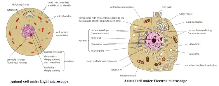
Compare the two diagrams given below. Name the structures in an animal cell that can be seen with the electron microscope but not with the light microscope.

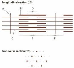
LSs and TSs of striated muscle fibres were examined in an electron microscope. In the given picture shows drawings of the structures visible in a sarcomere in LS and TS as seen in a TEM. Explain why an electron microscope rather than a light microscope was used to study these sections.
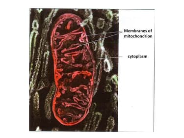
The mitochondrion in the figure given below is been magnified times. Calculate the real size of the image in and estimate the number of such mitochondrion that can be lined up to mark on a ruler.

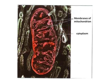
The mitochondrion given in the figure below is magnified times. Using a ruler, measure the length of the organelle and also calculate the real size of the image in .


